Introduction
A silastic drain is often inserted at the surgical site after reduction of facial bone fractures to minimize the formation of hematoma. But if not removed adequately, these drains may act as foreign bodies and induce chronic granulomatous wound [1,2,3,4,5]. These chronic granulomatous wound may be misdiagnosed as other lesion, such as abscess, epidermal cyst, or other neoplasm. The authors have experienced a case of a chronic granulomatous wound treated with wide excision and primary closure, and report our experience.
Case illustration
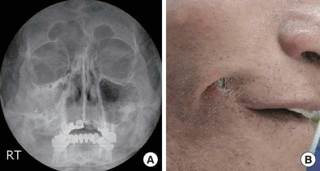
A 61-year-old man with a past history of facial bone fracture presented to our hospital with a progressive dimpling on his right cheek. Ten years ago, he underwent open reduction and internal fixation of the tripod fracture. Other than this surgery, he had no other medical or traumatic history. However, 3 months ago a painless firm nodule (1 cm in diameter) appeared beneath the skin of the right cheek that enlarged with time. The nodule was primarily diagnosed as an epidermal cyst in a local dermatologic clinic, and the patient was put on antibiotics, but showed little improvement. He was referred to our plastic surgery department due to the worsening of the skin wound accompanied by local heating and pain. On radiologic examination, a 5 cm linear radio-opaque material was seen, with underlying titanium plates and screws (Figure 1A).
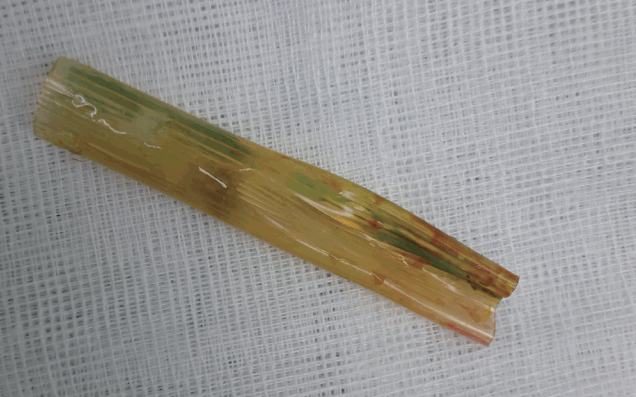
During the process of general anesthesia, pressure to the wound revealed the tip of silastic drain (Figure 1B). On careful dissection, it was apparent that the origin of the wound was from the silastic drain. It is guessed that the drain was not removed at the 10 years ago surgery (Figure 2). The granulomatous wound was widely excised and closed primarily. Antibiotics (cefazoline) for common skin bacterial pathogens were administered. Subsequent follow-up of the patient for 6 months revealed no complications and a good healing of the wound.
Discussion
The use of silastic drains after facial bone surgery is an old and well-established technique for the prevention of hematomas. Generally, these drains are removed within a week to prevent ascending infection. Because of its radiolucency, identification of a silastic drain on the radiograph is often difficult. In case the silastic drain has been misplaced, complete removal of the drain is essential for the prevention of infections.
Laxenaire et al. [6] found from his cohort of 137 patients that fibrous capsule formation, fistulas, and infections may be associated with silicone implantation. Another study has documented late extrusion of silicone implants through the facial skin at 10, 16, and 17 years after surgery [7].
Foreign body granulomatous reactions develop after a variable period of time ranging from 5 months to 15 years [8,9]. The clinical manifestations may vary from simple swelling, hardening, and tenderness to mild inflammation, burning sensations, and even fibrous reactions [10,11].
Complications of periorbital reconstruction are numerous and we may classify them as orbital or sinus complications, depending on the site of the inflammatory process. Among orbital complications, periorbit cellulitis, dacryocystitis, and diplopia are described, and several cases of cystic neoformation surrounding silastic sheet are reported [12,13].
The most common sinus complication is sinusitis, associated to rhinitis and purulent discharge; in literature, a case of bilateral sinusitis is described due to the silastic sheet perforation of the bony nasal septum and migration to the contralateral nasal cavity [14].
Chronic granulomatous wound forming after periorbital reconstructions is not common. This case reminds the surgeon that radiologic examination of a chronic skin lesion is essential for diagnosis, and that any misplaced silastic drains must be removed after operation.


