Introduction
Sunburn is the transient inflammatory response of skin, with symptoms such as mild erythema, pain, edema and blistering [1]. Most sunburns are first-degree or superficial second-degree, so there is generally no specific treatment other than symptomatic relief. While the symptoms of such sunburns usually resolve in 3 to 7 days, deep second-degree sunburns have been reported from time to time. Deep second-degree sunburns with extensive blistering, severe pain, and systemic symptoms may require hospitalization. The rate of disease progression varies because each individual has different levels of susceptibility to sunburn. Factors that can affect progression include the minimal erythema dose (MED), skin type, underlying disease, drug use, etc. [2]. We report a case of sunburn that deteriorated to a deep second-degree sunburn without a specific known factor in an otherwise healthy young female. The study was approved by the Institutional Review Board of Wonkwang University Hospital (IRB No. 2019-08-054) and performed in accordance with the principles of the Declaration of Helsinki. Written informed consent was obtained.
Case
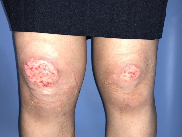
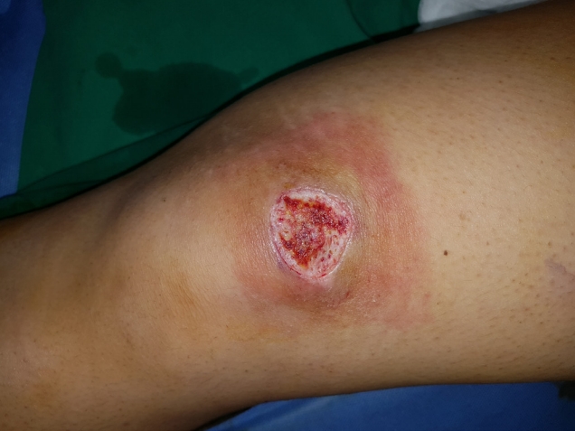
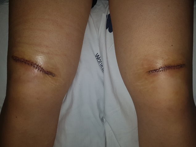
A 25-year-old female patient was sent to our hospital from a local clinic because of sunburn wounds on both knees. On the day of her injury, she wore black skinny jeans (97% cotton, 3% spandex) and watched a soccer game outdoors at around 2 PM for about an hour. The maximum temperature of that day was 28.2°C. The patient reported that it was overcast at the time of the game. The patient found blisters on both knees in the evening and visited the local clinic the next day. According to the local clinic, blister formation, including in the dermis, was observed, and a silver sulfadiazine dressing was applied after aspiration. The next day, because blisters had formed again, they were removed, and a silver sulfadiazine dressing was maintained. As the wound gradually dried, the dressing method was changed to a foam dressing. However, the patient did not improve, and was transferred to our hospital. At initial admission, upon taking patient history and physical examination, she was evaluated to have Fitzpatrick skin type III, had no underlying disease except anemia, and had no history of drug use except applied prednicarbate for urticaria on the neck 2 weeks prior. When she first visited (post-burn day 8), the wound was covered with a dry yellow eschar (Fig. 1), and we applied a hydrocolloid dressing to maintain the wet environment to remove eschar by promoting autolysis and also to inhibit bacterial growth by lowering the pH of the wound. As the wound status did not improve after 7 days with the hydrocolloid dressings, the decision was made to perform surgery. Because the patient was a young female who would wear skirts that exposed her knees, we elected to perform a flap operation instead of a skin graft. During the operation, we were able to confirm that the burn had progressed to the fat layer (Fig. 2). After debridement, we undermined both flaps at the fat layer level and the defect was covered with flap advancement (Fig. 3). There were no specific findings from the biopsy. At the 6-week follow-up visit the patient had recovered without complications, and received scar management and fractional laser abrasion treatments (Fig. 4).
Discussion
Sunburn is caused by excessive exposure to ultraviolet radiation (UVR) from sunlight, tanning, or phototherapy. The UVR is mostly ultraviolet A (UVA; 315–400 nm) and ultraviolet B (UVB; 280–315 nm). UVB contributes over 80% of the erythemally effective energy and has the most effective wavelengths to cause sunburn [2,3]. Therefore, when the UVB to UVA ratio is higher, the risk of sunburn is increased. The UVB to UVA ratio depends on latitude, season, and time of day. It is measured to be higher between 11 AM and 3 PM in summer [2]. The amount of cloud cover is also an important factor. Though cloud provides some protection, caution should be taken because significant quantities of UVR reach the earth’s surface even in cloudy weather [4]. As for known risk factors, there is alcohol consumption, smoking, body mass index, physical activity, sleep quality, fruit intake, altitude and reflection [5-7].
Because the susceptibility to sunburn varies in each person, the course of the disease also differs by person. The individual’s susceptibility to sunburn is clinically important because it serves as a risk marker for melanoma and nonmelanoma skin cancer [8,9]. Susceptibility to sunburn can be assessed by determining the MED, widely understood as the threshold dose for sunburn, or the lowest dose that produces erythema within 1 to 6 hours and disappears within 24 hours. The susceptibility to sunburn also varies according to the individual’s skin type [2]. The Fitzpatrick skin phototyping scale classifies skin into six groups according to skin color and the skin’s reaction after sun exposure [10]. MED also differs by skin type [11].
The diagnosis of sunburn is based on clinical findings and patient history. For clinical findings, the degree of pain and erythema and whether blistering exists are the basis of a diagnosis. History and evaluation include the duration, area, and degree of exposure and whether there was any history of photosensitizing drugs or topical photosensitizer use, phototherapy, or tanning. Photosensitizing drugs reduce the dose threshold for erythema and typically include anticancer drugs, antidepressants, antimicrobials, antiparasitic drugs, antipsychotic drugs, diuretics, hypoglycemic agents, nonsteroidal anti-inflammatory drugs, antihistamines, antihypertensive drugs, and oral contraceptives [12]. However, we did not believe the prednicarbate ointment used by the patient was a cause because prednicarbate is not one of the photosensitizing drugs listed above, and the patient had used the ointment intermittently for only 1 week after the onset of symptoms.
Jackson [13] introduced the three-zone theory of burn wounds in 1953. The zone of coagulation is located at the center of the wound and characterized by irreversible tissue loss. However, the surrounding zone of stasis is characterized by reduced tissue perfusion and a mixture of viable and nonviable tissue. Thus, if proper wound management is performed, reversible pathophysiological changes may occur. The primary tissue injury involves protein denaturation and loss of plasma membrane integrity, which can be caused by heat, chemicals, electricity, friction, ultraviolet, and freezing. The loss of normal skin barrier function leads to burn injury complications [14]. As we could not find any sunburn wounds in areas exposed to sunlight that required treatment other than the patient’s knees, we believed there were other factors that had deteriorated the wound. The authors suggest that the patient’s primary tissue injury was due to repetitive friction in the knee area from the skinny jeans worn by the patient when she watched the game, with UVR inducing a second-degree wound with vesiculation. In addition, the initial treatment of a 1% silver sulfadiazine dressing may have interfered with the wet environment of the wound. But it is difficult to diagnose it as the sole cause of the progression of the disease [15]. We suggest that the lesion expanded to the surrounding zone of stasis because of inappropriate wound care management. This underscores the importance of understanding the pathophysiologic responses after a burn injury to ensure a better clinical outcome.
This case showed that sunburn can proceed to a deep second-degree without a specific known factor, and that sunburn can occur in ordinary early summer weather, even when the area is clothed. Though most sunburns are treated as first-degree or superficial second-degree, there are some deep seconddegree sunburns occasionally reported. As mentioned above, there are many factors that may contribute to the progression of the deep second-degree sunburn, but as in this case, such sunburns can occur even though the known key risk factors were not involved. We therefore need to examine sunburns and observe their initial treatment with more caution. In addition, more care should be given to prevent sunburn, including staying out of the sun, wearing protective and well-ventilated clothing, avoiding wearing tight clothing, and using sunscreen.