Introduction
Though physicians suspect Marjolin’s ulcer in cases of chronic skin ulcerations, including those found in areas of post-burn scar contracture [1], a condition not to be mistaken as Marjolin’s ulcer is heterotopic ossification (HO). HO is caused by the formation of extraskeletal bone from calcium abnormally deposited in soft tissues, but its underlying cause is not known. HO usually occurs in burn patients or after hip arthroplasty, fracture, dislocation, or general paralysis [2], and is usually found in muscle layers subjected to trauma or surgical sites and around joints [3]. This report is to discuss the cause, clinical character, and the treatment method used for a case of unusual skin ulceration initially suspected as skin cancer, which was actually caused by HO of subcutaneous tissue in the patient’s thigh. The study was performed in accordance with the principles of the Declaration of Helsinki. The informed consent was waived.
Case
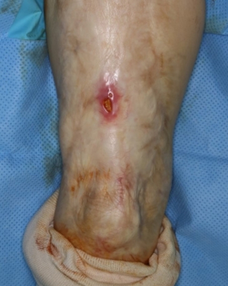
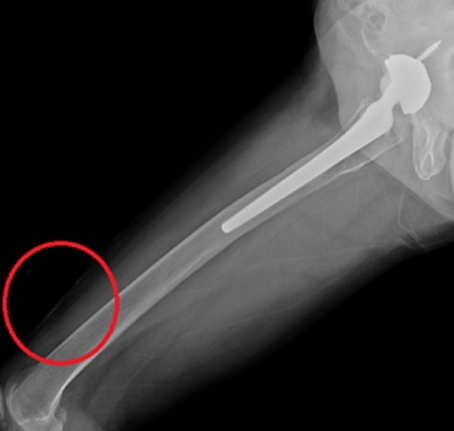
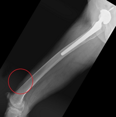
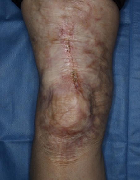
A 68-year-old woman who had burnt her right thigh 50 years previously and had undergone hip joint hemiarthroplasty 10 years ago visited our department with a 2-year-old skin ulceration, 1 cm in diameter, on her right anterior thigh (Fig. 1). We suspected Marjolin’s ulcer and performed a biopsy and a routine X-ray. Histopathology showed fibrosis and calcification of the subcutaneous fat layer, accompanied by chronic inflammation but no evidence of skin cancer. The X-ray revealed multiple linear bony calcifications in subcutaneous tissues (Figs. 2, 3), but no calcifications in the hip joints or muscle. Serum calcium, phosphorus, and alkaline phosphatase levels were within normal ranges (calcium, 9.4 mg/dL; phosphorus, 3.5 mg/dL; alkaline phosphatase, 83 IU/L). We diagnosed skin ulceration due to HO of subcutaneous tissue, and excised the skin ulceration along with the heterotopic calcification. The excised wound was closed with Prolene 4-0 and nylon 5-0. The wound healing process was uneventful (Figs. 4, 5). After the operation, she had no calcification or ulceration on her thigh for 11 months.
Discussion
Marjolin’s ulcer is an aggressive ulcerating form of squamous cell carcinoma that presents in areas previously traumatized or in chronically inflamed or scarred skin [1]. When Marjolin’s ulcer is suspected, biopsy, computed tomography, and magnetic resonance imaging are usually performed for accurate diagnosis and to also to assess the extent of the tumor.
Meanwhile, HO mostly occurs after a severe burn or trauma [4]. HO associated with previous burn injury, fracture, dislocation, or limb paralysis takes place around joints and muscles, and is caused by abnormal bone morphogenic protein (BMP) expression in injured muscle. Conditions that precede the onset of HO include trauma, hematoma, signals arising from inflammatory reactions at sites of injury, and an environment conducive to HO, such as immobilization and extended bed rest.
Furthermore, the incidence and severity of HO is related to the extent and location of a burn, how long movement had been limited, the degree and duration of paralysis, the time taken for wound healing, the presence of infection, hemorrhage, hypoxia, and the degree of post-traumatic hematoma [5,6]. Fibrodysplasia ossificans progressiva and traumatic myositis ossificans are also viewed as types of HO.
Symptoms of HO, which is usually observed around large joints and muscle layers, include joint stiffness, joint dyskinesia, edema, pain, and dysfunction of the affected area. This type of ossification is found incidentally on radiographs. In the absence of symptoms, no special treatment is required, but when the joint is in pain, swelling, and motion is limited, it must be treated. Metabolic features of HO include normal calcium and phosphorus levels in serum. Elevated serum alkaline phosphatase levels observed at the initiation of bone formation are followed by a return to normal levels [7]. Prophylactic treatment including physical therapy can be performed if a patient is immobilized for an extended period of time due to neurologic causes, old age, or severe trauma, or when severe ossification is expected due to severe injury. Nonsteroidal antiinflammatory drugs and radiation therapy are suggested nonsurgical treatment methods. However, HO in the subcutaneous tissue layer is not associated with joint symptoms or stiffness. Instead, patients visit the hospital when there are symptoms such as a nonhealing ulcer, as in our case. When diagnosis and treatment are delayed for a protracted period the risk of malignant transformation is substantially increased [8], and when HO limits joint movement, surgical removal of ossifications around joints is mandatory [3].
Several hypotheses have been proposed to explain the etiology of HO after trauma. Some physicians believe primitive mesenchymal cells may transform into osteogenic cells after trauma. Studies continue to show external stimuli result in the expression of osteogenic precursor cells and HO, and that BMP plays an important role in this process. Several subtypes of osteogenic factors, that is, BMP 2, 4, 6, 7, and 9, regulate osteoblast regulation and bone formation [9]. Two types of cells are known to be involved in HO within the muscle, namely myogenic stem cells or satellite cells, and muscle-resident mesenchymal or stromal cells, which are present around connective tissues.
In cases of chronic skin ulceration, surgeons should suspect malignancies such as Marjolin’s ulcer and squamous cell carcinoma. However, as skin ulcerations are not always skin cancer, it is imperative for clinicians to evaluate the routine X-ray and preoperative biopsy and remember to look for unusual skin lesion such as an unusual heterotopic calcification on postburn scar contractures.